
Veterinary-clinical pathology
Veterinary-clinical pathology is a branch of pathology and a special veterinary-medical field. The diagnosis of various diseases is carried out by means of laboratory analyses of bodily fluids and cells. The areas include hematology, biochemistry, hormones, cytology and laboratory general management.
The platform www.e-fur.at
… is a veterinary-clinical-pathological laboratory service and is available to all vet colleagues in diagnostic enquiries regarding:
- Cytology
- Blood interpretations
- Laboratory diagnostic-further recommendations of various tests
- Support for sample acceptance
- Installation, optimization and management of your in-house laboratory.
In order to save precious time, the services are available digitally, where practically executable. Sending slides e.g. by post is of course also possible.
- Upload static images of cytological preparations using a mobile phone camera or microscope camera in the “portal”. Digitally fill in the request sheet and press send.
- Upload blood findings in the “portal”, fill in the request form digitally and send them.
- Written findings are usually received on the same day. In urgent cases, even every minute.
- Please take details in “Services”.
For further questions, please leave a message in the “Contact” button. I look forward to working together!
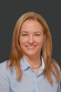
Erika Furman dr.vet.med., Diplomate ECVCP
EBVS® European Specialist in Veterinary Clinical Pathology
About the person
I was born in Fürth, Germany, where I lived with my family until I was eight years old. Through my move to Slovenia I completed the primary school and the high school there. I also studied veterinary medicine in Slovenia (Veterinary Faculty of the University of Ljubljana).
I first enjoyed specialist-level clinical pathology during a two-week working visit to the UK at Dick White Referrals in January 2010. There I was recommended for a residency at the European College for Veterinary Clinical Pathology (ECVCP), which I successfully completed at Labor InVitro Vienna (now Idexx) from 2011 to 2015.
I passed the examination to become a Diplomate ECVCP, the European Specialist in Veterinary Clinical Pathology, in 2017. At InVitro/Idexx in Vienna, I worked as a senior clinical pathologist and deputy manager until January 2019.
Before my “laboratory time” I worked for 14 years in two small animal clinics, Toplica in Slovenia and veterinary clinic Hollabrunn in Austria. My work areas were diverse, such as routine patient examinations, anesthesia, diagnostic oncology and oncological care of patients including chemotherapy, intensive medicine, etc. But here, too, my preferences were of cytology and laboratory diagnostics.
With the foundation of the platform www.e-fur.at, the wonderful diagnosis of clinical pathology at specialist level is quickly accessible to all colleagues. Only one “mouse click” removed.
Erika Furman dr.vet.med., Diplomate ECVCP
EBVS® European Specialist in Veterinary Clinical Pathology
Publications
Cytology
The cytological examination of various tissue structures and fluids is a relatively simple, cost-effective, rapid and established examination method. A final diagnosis is not always achieved, but it is often possible to successfully use cytology as a guide for further diagnostics. The production of diagnostic samples is crucial for a targeted cytological diagnosis.
With existing expertise, the evaluation can be carried out in practice. Sending to a veterinary laboratory with specialists in cytology is the other choice. Telecytology, also known as digital cytology or digital microscopy, is a new and so far the fastest way to send samples to the cytologist. For this, the slide must be colored and digitized.
A distinction is made between all-slide and static digitization.
Full-object object digitization can be carried out by means of microscope cameras or scanning cameras, but of course where the devices are also available. Static digitization, which is very widespread, deals with static recordings of regions of interest (region of interest; ROI), which can be made with the help of mobile phone cameras.
You need a steady hand or certain adapters (e.g. Besser De-Luxe Smartphone Adapter) to clamp and fix the phone to the eyepiece. Static images of ROIs are easier to take using microscope cameras. The evaluation is based on the recordings, for which a high-quality production of ROIs of cytological slides is of extreme importance.
I investigate cytological cases that arrive at me digitally or by classical submission via post or messenger.
Literature:
- Blanchet et al. Evaluation of Region of Interest Digital Cytology Compared to Light Microscopy for Veterinary Medicine. Vet Pathol. 2019 May 21.
- Bertram et al. The pathologist 0: an update on digital pathology in veterinary medicine. Vet Pathol. 2017;54(5):756–766.
- Bertram et al. Validation of digital microscopy compared with light microscopy for diagnosis of canine cutaneous tumors. Vet Pathol. 2018;55(4):490–450.
- Maiolino et al. Evaluation of static telepathology in veterinary diagnostic cytology. Vet Clin Pathol. 2006;35:303–306.
Interpretation of findings
Findings of various blood tests such as hematology, biochemistry or endocrinology, which were measured in-house or by submission laboratories, can be sent to me for an interpretation. Of course, urinary findings, cytology findings and/or blood smears are also possible. Then I create a written evaluation of all deviations, a so-called clinical-pathology report.
You will receive a written interpretation of the findings with a diagnosis or at least a probability diagnosis, differential diagnoses and recommended further diagnostics with an emphasis on laboratory medicine.
In the “Portal” you can easily upload all relevant findings, send them to me, fill in the enclosed request form and add certain questions.
In-house laboratory optimization
Every colleague needs a high-quality, fast and reliable laboratory diagnostics for his work.
I evaluate your in-house laboratory, make suggestions for improvements and fight with the analyzers in case of problems. Setting up analyzers, validations, quality control, reference intervals, calculations of laboratory costs and much more are also some of my strengths.
If necessary, please contact me via “Contact” with a short description of the problem.
Contact
The entered data will only be processed for the purpose of processing your request. Further information can be found in our privacy policy.